Plantar fasciitis is one of the most common musculoskeletal complaints seen in clinical practice — and one of the most commonly mismanaged. The standard narrative goes something like this: rest it, stretch your calf, get better insoles, maybe get a cortisone shot. For some people, this approach works. For many others, the pain returns, becomes chronic, or never fully resolves.
The reason is that plantar fasciitis is rarely just a problem with the plantar fascia. It is a whole-chain condition that expresses itself at the heel but is frequently driven by dysfunction well above it. Understanding that bigger picture is the difference between temporary relief and lasting recovery.
Accurate Diagnosis: Starting at the Heel
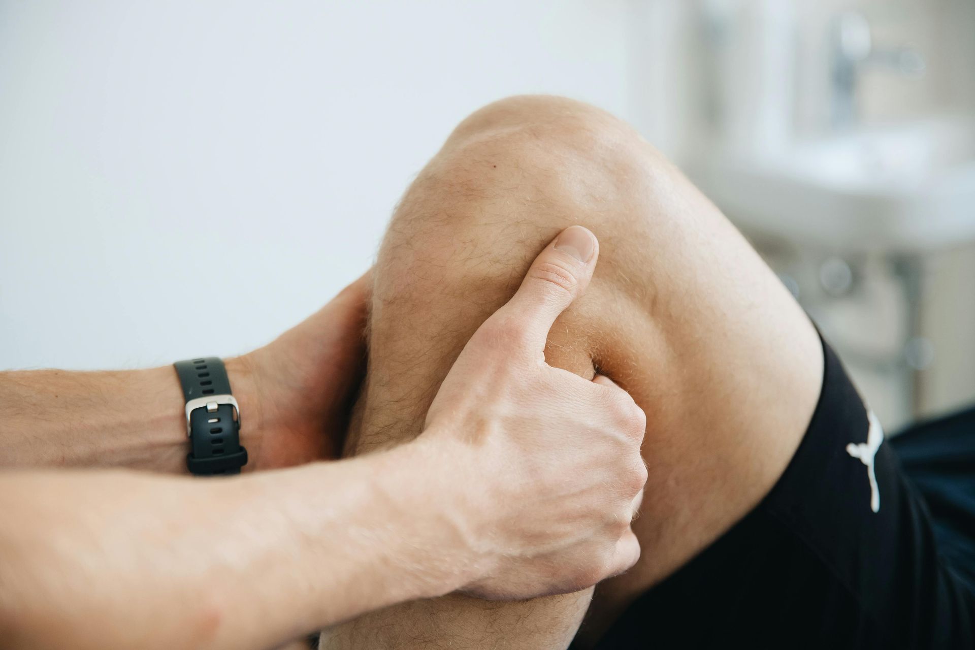
Before anything else, the diagnosis needs to be confirmed. The gold standard for diagnosing plantar fasciitis is the reproduction of the patient's familiar pain with firm palpation at the medial calcaneal tubercle — the bony prominence at the inside of the heel where the plantar fascia attaches.
This specificity matters because plantar foot pain has several possible origins that are frequently confused with plantar fasciitis and require different treatment. The differential diagnosis includes stress fractures of the calcaneus, plantar nerve entrapment, neuromas, and S1 nerve root radiculopathy from the lumbar spine. If pain cannot be consistently reproduced by palpation at the medial calcaneal tubercle, it may be plantar pain — but it may not be plantar fasciitis, and treating it as such will produce limited results.
The Proximal Chain: Looking Upstream
Once the local diagnosis is confirmed, the more important clinical question is: what is driving it? The research increasingly points to proximal chain dysfunction as a primary contributor to plantar fasciitis — meaning that weakness, stiffness, or altered mechanics in the thigh, hip, and lumbar spine are loading the plantar fascia abnormally from above.
Key upstream contributors to investigate include:
- Hamstring and quadriceps weakness: The posterior chain plays a critical role in shock absorption and force distribution during walking and running. Weakness here shifts load to the foot and ankle.
- Hip mobility deficits: Restricted hip extension changes the mechanics of the push-off phase of gait, increasing plantar fascia strain with every step.
- Thoracic and lumbar hypomobility: Stiffness in the mid and lower back alters walking mechanics in ways that accumulate force at the foot over thousands of steps per day.
- L5-S1 nerve root involvement: The nerve roots at this level innervate the gluteus maximus and medius — key hip stabilizers. Compromise here creates hip weakness that drives compensatory foot mechanics.
Addressing only the foot while ignoring these contributors is one of the primary reasons plantar fasciitis recurs. The foot is the symptom. The chain is the cause.
Local Treatment: What Works at the Foot
Effective local treatment addresses tissue quality, joint mobility, and the neural environment at the foot and ankle:
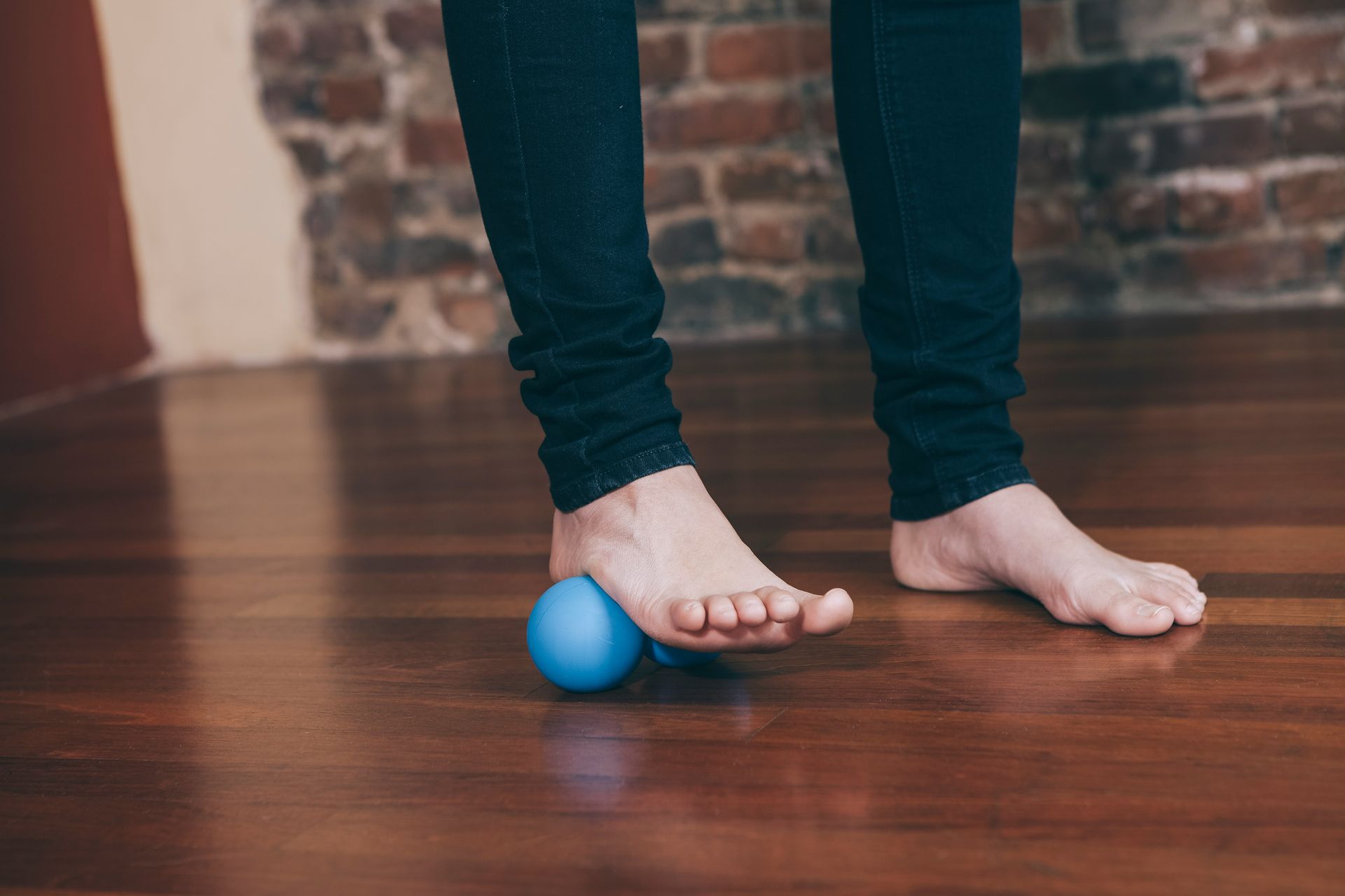
- Instrument-Assisted Soft Tissue Mobilization (IASTM): Targeted work to the gastrocnemius, soleus, and anterior tibialis reduces tissue restriction, improves blood flow, and addresses the chronic tensile load that these muscles place on the plantar fascia through the Achilles and heel.
- Dry needling: Targeting the adductor hallucis and quadratus plantae at their attachment to the medial calcaneal tubercle, along with trigger points in the medial gastrocnemius, can produce significant and rapid pain reduction.
- Joint mobilization: Restoring talocrural (ankle) and hip joint mobility ensures that the foot and lower extremity have the range of motion necessary for normal gait mechanics — reducing the compensatory strain on the plantar fascia.
Loading the Tissue: The Strengthening Progression
Passive treatment alone does not resolve plantar fasciitis. The plantar fascia and the calf-Achilles complex need progressive loading to remodel, strengthen, and adapt. The loading progression follows a logical sequence:
- Seated heel raises: Begin with the seated position to isolate the soleus and load the plantar fascia with reduced body weight. Focus on full range of motion and controlled tempo.
- Standing calf raises — V position: Standing with heels together and toes angled outward, emphasizing push-through the big toe. This recruits the peroneus longus and prevents the common compensation of rolling to the outer foot. Use 50/50 weight distribution between feet.
- Single-leg calf raises on a step: With the heel hanging off the edge of a step, perform full-range eccentric-concentric calf raises. The eccentric (lowering) phase is the most therapeutically important component and should be slow and controlled.
- Progressive loading: As tolerance develops, add load via a weighted vest or dumbbell. Monitor both the quantity and quality of each repetition — form deterioration is a signal to stop, not push through.
Addressing the Hip and Posterior Chain
Corrective exercises targeting the proximal chain are essential for preventing recurrence and should run in parallel with local foot treatment:
- Clamshell protocol: Side-lying with knees bent at 90 degrees, rotating the top knee upward without allowing the pelvis to roll back. 2-3 sets of 15-20 reps. Targets the gluteus medius and improves hip stability.
- Bridge to single-leg bridge progression: Standard glute bridge first, progressing to single-leg as strength allows. 2-3 sets of 12-15 reps. Addresses posterior chain weakness and improves hip extension mechanics.
- Single-leg deadlift: Standing on one leg, hinging at the hip with the free leg extending behind. 2-3 sets of 10-12 reps per side. Trains hamstring strength and eccentric control through the full range — directly relevant to the push-off phase of gait.
- Standing hinge pattern loading: Progressing through deadlift variations, step-ups, and hip thrusters as strength develops. These movements replicate the mechanics of walking and running and are the endpoint of the rehabilitation progression.
The Thomas Test: Assessing Hip Flexor Contribution
One assessment worth understanding is the Thomas test, which evaluates tightness in the hip flexors and quadriceps. In patients with plantar fasciitis, compensatory shortening of the hip flexors and rectus femoris is common — a consequence of the altered gait mechanics that develop alongside chronic heel pain. Tightness here contributes to reduced hip extension, which in turn increases plantar fascia loading during walking.
When hip flexor restriction is identified, the Thomas pump exercise — a therapeutic technique performed with the patient supine and the affected leg hanging off the table edge — eccentrically lengthens both the hip flexors and quadriceps while simultaneously activating the glutes through reciprocal inhibition. It is a highly efficient way to address the hip flexor component of the condition.
Timeline and Expectations
With a comprehensive approach — accurate diagnosis, local treatment, progressive loading, and proximal chain correction — most patients with plantar fasciitis experience significant improvement within 6-12 weeks. Chronic cases that have been present for more than a year may require longer timelines, but the same principles apply.
The key mindset shift is moving from passive management (rest, ice, insoles) to active rehabilitation. The plantar fascia is connective tissue — it remodels in response to progressive load. Rest alone does not remodel it. Targeted loading does.
Plantar fasciitis that has not responded to conventional treatment is almost always a signal that the full chain has not been assessed and addressed. At Performance Collective, we look at the foot in the context of everything above it — because that is where durable resolution comes from.
 RAD Roller on Unsplash
" id="1270948247" class="" data-dm-image-path="https://irp.cdn-website.com/1b899acb/dms3rep/multi/rad-roller-FOhZ2ZMwr1I-unsplash.jpg" width="1920" height="1280" onerror="handleImageLoadError(this)"/>
RAD Roller on Unsplash
" id="1270948247" class="" data-dm-image-path="https://irp.cdn-website.com/1b899acb/dms3rep/multi/rad-roller-FOhZ2ZMwr1I-unsplash.jpg" width="1920" height="1280" onerror="handleImageLoadError(this)"/>